
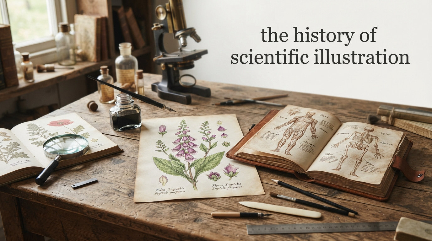
Scientific illustration stands at the unique intersection of art and science, where detailed drawings have documented everything from the tiniest cells to the vastness of cosmic phenomena. The history of scientific illustration spans thousands of years, evolving alongside human understanding of the natural world. Before photography existed, skilled illustrators served as the primary means of recording and communicating scientific discoveries. These artists combined artistic talent with deep anatomical and observational knowledge to create images that shaped how generations understood science. Today, scientific illustration continues to play a vital role in research, education, and public outreach, even as digital tools transform the field.
Table of Contents
What is Scientific Illustration?
Scientific illustration is the practice of creating detailed artwork to visually communicate scientific concepts, specimens, and discoveries. Unlike general art, scientific illustrations must achieve absolute accuracy while remaining clear enough to convey complex information to viewers. Illustrators often work closely with researchers, studying specimens through microscopes and performing their own dissections to understand structures before rendering them. The tradition encompasses many specialized fields, including botanical illustration, zoological illustration, anatomical illustration, and entomological illustration. Each discipline demands its own set of observational skills and technical approaches to capture subjects truthfully.
Throughout the history of scientific illustration, artists have served as essential collaborators to researchers and naturalists. Their work appeared in textbooks, scholarly publications, museum displays, and field guides, making scientific knowledge accessible to wider audiences. The best scientific illustrations achieve what photographs often cannot: they highlight the most important features of a specimen while eliminating visual noise and distortion. Illustrators can also show multiple views, cross-sections, and developmental stages in a single image, providing information that a single photograph cannot convey. This ability to synthesize and clarify makes scientific illustration an indispensable tool for education and research communication.
Prehistoric Origins of Scientific Illustration
The story of scientific illustration begins in the depths of prehistory, with cave paintings that represent some of humanity’s earliest attempts to document the natural world. Cave paintings in Lascaux, France, and Altamira, Spain, dating back approximately 17,000 years, depict animals with remarkable anatomical accuracy. These ancient artists understood the skeletal structure and musculature of their subjects, evident in how the animals’ legs and bodies are rendered. The cave paintings at Chauvet, estimated to be over 30,000 years old, show an even more sophisticated understanding of animal forms. Paleolithic artists depicted mammoths, horses, cattle, and deer with a fidelity that suggests direct observation of these animals in their habitats.
Archaeological evidence from these sites shows that prehistoric artists used natural pigments derived from iron oxides, manganese, and charcoal to create their images. The application techniques varied from finger painting to spraying pigment through hollow bones, demonstrating early experimentation with different tools and methods. Some researchers believe the artists may have used animal fat as a binding agent for the pigments, which would have helped the colors adhere to the rough cave surfaces. The preservation of these paintings over millennia provides invaluable insight into the wildlife that existed during the Upper Paleolithic period.
Rock art found across Africa, Australia, and the Americas similarly shows careful observation of plants and animals in an artist’s environment. Indigenous Australian rock art includes detailed depictions of local flora and fauna that served both cultural and practical purposes. Australian Aboriginal rock art sites contain images of extinct megafauna like the giant kangaroo and the marsupial lion, providing scientific data alongside cultural significance. Many prehistoric illustrations appear to combine documentation with spiritual or ceremonial significance, suggesting that even ancient humans understood the power of visual representation for conveying knowledge. The accuracy of these early works reveals a deep engagement with the natural world that forms the foundation of scientific observation.
Scientific Illustration in Ancient Civilizations
Ancient Egyptian civilization advanced scientific illustration significantly, with detailed paintings and carvings depicting plants, animals, and human anatomy. Egyptian tomb paintings show sophisticated knowledge of anatomy, particularly in scenes showing surgical procedures or the preparation of bodies for mummification. The Edwin Smith Papyrus, dating to around 1600 BCE, contains illustrations of anatomical features alongside hieroglyphic text describing treatments and diagnoses for wounds and injuries. These documents reveal that the Egyptians distinguished between different types of wounds and had developed systematic approaches to medical documentation.
The Egyptians depicted hundreds of plant and animal species in their art, often with enough accuracy for modern biologists to identify specific species. Botanical representations in tomb paintings show detailed renderings of lotus flowers, papyrus reeds, and various trees that grew along the Nile. Zoological illustrations included detailed depictions of cattle, hippos, crocodiles, and numerous bird species, showing careful observation of animal behavior and physical characteristics. Egyptian artists also created detailed maps and plans for architectural and construction purposes, representing an early form of technical drawing that served practical purposes.
Greek scholars further developed scientific illustration, with artists accompanying physicians like Hippocrates to document anatomy and medical conditions. Aristotle’s works on biology included observations of animals that were sometimes accompanied by drawings, establishing a tradition of combining textual description with visual representation. Greek naturalists compiled encyclopedic works that included illustrations of animals and plants from across the Mediterranean world. The Greeks also produced early botanical illustrations, with manuscripts of Dioscorides’s herbal guides dating to the 1st century CE containing carefully rendered plant images that remained influential for centuries.
Roman naturalists like Pliny the Elder compiled encyclopedic works that included illustrations of animals and plants from across the known world, though many of the original images have been lost to history. Roman frescoes from Pompeii include remarkable depictions of garden plants and wildlife, showing the continued integration of scientific observation and artistic representation in daily life. The Romans also developed maps and topographical drawings for military and administrative purposes, representing practical applications of illustration techniques.
Medieval Era: Manuscripts and Early Documentation
During the medieval period, scientific knowledge and illustration were preserved primarily within monastery scriptoriums, where monks copied and illuminated ancient texts. Botanical manuscripts called herbals featured hand-painted illustrations of medicinal plants, often with accompanying text describing their properties and uses. These illustrations varied widely in accuracy, with some showing remarkable detail while others were highly stylized or symbolic. Monks like those at the Abbey of Saint Gall created elaborate manuscript traditions that preserved classical knowledge while adding their own observations of local plants and animals.
Medical manuscripts during the medieval period often included anatomical diagrams that reflected the prevailing understanding of the human body, influenced heavily by Galenic anatomy. These diagrams typically showed the body as a series of interconnected tubes and vessels, reflecting the importance placed on bloodletting and humoral theory in medieval medicine. Despite their inaccuracies, medieval anatomical illustrations served an important function in preserving and transmitting medical knowledge across generations of practitioners. Monastic manuscripts also included bestiaries, illustrated collections of animals that combined natural history with moral and religious teachings.
Islamic scholars during the Golden Age made significant contributions to scientific illustration, preserving and expanding upon classical knowledge. Persian and Arabic manuscripts include detailed illustrations of astronomical instruments, anatomical diagrams, and botanical specimens that often surpassed their classical predecessors in accuracy and detail. The Book of Animals by al-Jahiz and works by Ibn al-Haytham on optics included sophisticated diagrams showing the principles being discussed. Islamic scholars also developed more systematic approaches to illustration, emphasizing accuracy and clarity in representing scientific concepts.
The tradition of Islamic scientific illustration included detailed star charts and astronomical instruments that reflected the advanced state of Islamic astronomy during the medieval period. Scholars like Al-Biruni created detailed geographical maps and topographical drawings that demonstrated sophisticated understanding of spatial relationships and measurement. Islamic medical texts included carefully rendered anatomical illustrations that preserved and expanded upon Greek and Roman traditions. These manuscripts spread throughout Europe and the Middle East, influencing the development of scientific illustration in subsequent centuries.
The Renaissance Revolution in Scientific Illustration
The Renaissance transformed scientific illustration from a primarily decorative art into a powerful tool for understanding the natural world. The development of the printing press around 1440 allowed illustrations to be reproduced and distributed on an unprecedented scale, spreading scientific knowledge throughout Europe. The combination of printed text and images enabled new forms of scientific communication that were faster and more accurate than previous manuscript traditions. Woodcut and copperplate engraving techniques improved dramatically, allowing for finer detail in printed illustrations than had previously been possible.
This period saw the emergence of illustrated scientific publications that reached wider audiences than ever before, democratizing access to knowledge about the natural world. The availability of printed herbals, anatomical texts, and natural histories created new demand for accurate and aesthetically pleasing illustrations. Publishers actively recruited skilled artists to work with scientists and naturalists, establishing models of collaboration that persist today. The relationship between printing technology and illustration advancement created feedback loops that accelerated progress in both fields.
Leonardo da Vinci stands as perhaps the most famous figure in the history of scientific illustration, creating thousands of anatomical drawings that remain astonishing in their accuracy. Da Vinci performed numerous dissections to understand the human body, producing detailed studies of muscles, bones, and organs that surpassed anything previously attempted. His notebooks show observations from multiple angles, with cross-sections and layered views that reveal internal structures. Da Vinci combined artistic skill with scientific curiosity in ways that set new standards for the field.
Da Vinci’s studies extended beyond human anatomy to include comparative anatomy of animals, botanical studies of plants, and geological observations of rock formations and fossils. He created detailed drawings of the human heart, brain, and eye that anticipated discoveries made centuries later. His notebooks also contain studies of water flow, bird flight, and the movement of light, demonstrating the breadth of his scientific curiosity. Da Vinci believed firmly that observation and direct study of nature were essential for accurate representation, a principle that remains central to scientific illustration today.
Andreas Vesalius revolutionized anatomical illustration with his groundbreaking work “De Humani Corporis Fabrica,” published in 1543. Vesalius worked with skilled artists to create precise woodcut illustrations of human anatomy that corrected centuries of errors inherited from Galen. His illustrations showed bodies in active poses rather than the static figures common in earlier anatomical works, making them more useful for teaching and understanding. The Fabrica set a new standard for the integration of text and image in scientific publishing.
The quality of Vesalius’s illustrations was matched by the book’s sophisticated layout and printing, which allowed the images to work in concert with the written text. Each illustration was positioned to correspond with relevant passages, creating a seamless integration of word and image. The book included illustrations of the skeleton, muscles, nerves, blood vessels, and internal organs, providing the most comprehensive anatomical description of its time. Its influence extended throughout Europe, transforming how anatomy was taught and understood in medical schools across the continent.
Natural History and Exploration Era
The 17th and 18th centuries saw an explosion of interest in natural history, driven by European exploration and the discovery of new species around the world. Naturalists like Maria Sibylla Merian exemplified the spirit of careful observation that characterized this period. Merian traveled to Suriname in 1699 to study and document tropical insects and plants, producing stunning illustrations that combined scientific accuracy with artistic beauty. She was one of the first artists to show insects in their full life cycles, including the plants they fed upon.
Merian’s work was remarkable not only for its scientific accuracy but also for its ecological perspective, showing the relationships between different species in their natural habitats. Her illustrations documented metamorphosis in ways that were previously poorly understood, showing caterpillars transforming into butterflies and other dramatic life changes. Her dedication to observation extended to raising live insects in her studio to study their behavior and development. Her work demonstrated the importance of observing specimens in their natural environments rather than relying on preserved or incomplete samples.
Botanical illustration flourished during this period, with royal botanical gardens and private collectors commissioning detailed studies of plant species. Artists developed increasingly sophisticated techniques for depicting plant structures, including root systems, cross-sections of stems, and detailed studies of flowers and seeds. The development of binomial nomenclature by Carl Linnaeus created new demands for accurate illustrations that could distinguish between similar species. Illustration became essential for taxonomy, with plates showing key identifying features that could differentiate between species described in the new systematic framework.
Explorers and naturalists returning from voyages around the world brought back countless new species that required documentation and illustration. Scientific expeditions often included dedicated illustrators whose role was to record the plants and animals encountered during the journey. These illustrated records served both scientific and archival purposes, preserving information about species that might be lost as European expansion transformed ecosystems around the world. The specimens and illustrations collected during these voyages formed the basis for major scientific collections and publications that shaped the emerging discipline of natural history.
The 19th Century Golden Age of Scientific Illustration
The 19th century represents the golden age of scientific illustration, when elaborate printed images reached their peak of technical sophistication and artistic achievement. Advances in lithography allowed artists to create images with a subtlety and range previously impossible in print reproduction. Color printing techniques improved dramatically, enabling the mass production of stunning natural history volumes. Lavishly illustrated works on birds, shells, insects, and plants became available to wealthy collectors and institutions, democratizing access to visual scientific knowledge.
The publishing industry during this period saw the emergence of grand natural history projects that remain landmarks in the history of scientific illustration. These multi-volume works often took decades to complete and required teams of illustrators, engravers, and editors working in concert. The financial investment required for such projects meant that they were often sponsored by wealthy patrons, scientific societies, or governments interested in promoting natural knowledge. The competition among publishers to produce the most impressive illustrated works drove continuous improvements in printing and illustration techniques.
John James Audubon created one of the most famous works in the history of scientific illustration with “The Birds of America,” published between 1827 and 1838. Audubon’s life-sized illustrations of North American birds were remarkable for their size, detail, and naturalistic settings. He hunted, posed, and drew birds from fresh specimens, capturing their colors and postures with unprecedented accuracy. The resulting work contains 435 hand-colored, life-sized prints that remain unsurpassed in their combination of scientific rigor and artistic vision.
Audubon’s approach emphasized showing birds in their natural habitats, combining ornithological documentation with landscape painting in ways that influenced generations of subsequent illustrators. His close observation of bird behavior resulted in illustrations that captured not just the appearance but also the character and movement of living birds. The technical challenges he overcame in creating life-sized prints of such quality were enormous, requiring innovative printing methods and careful color matching. Audubon’s work established new standards for what natural history illustration could achieve.
Ernst Haeckel expanded the boundaries of scientific illustration with his detailed depictions of marine life, particularly the microscopic organisms he studied. His work “Kunstformen der Natur” (Art Forms in Nature), published between 1899 and 1904, featured 100 stunning plates showing radiolarians, medusae, and other creatures with extraordinary precision. Haeckel’s illustrations revealed the hidden beauty of microscopic life, showing geometric patterns and symmetries that fascinated scientists and artists alike.
Haeckel’s work influenced both scientific understanding and the Art Nouveau movement, demonstrating how scientific illustration could bridge traditionally separate worlds. His detailed renderings of single-celled organisms and other microscopic life forms helped make these invisible creatures accessible to wider audiences. Many of Haeckel’s illustrations remain visually striking today, more than a century after their creation. His systematic approach to documenting biological diversity helped establish the field of marine biology and inspired subsequent generations of illustrators.
Elizabeth Gould contributed significantly to ornithological illustration, producing hundreds of plates for works by her husband John Gould and others. She illustrated numerous species of birds from around the world, combining accuracy with an elegant artistic style. Despite working in an era when women were excluded from formal scientific institutions, Gould’s skill and dedication earned her recognition among naturalists. Her work on birds from Australia, Asia, and other regions documented species that were new to European science.
Other women illustrators made important contributions during this period, though their work was often attributed to male editors or publishers. Mary Louise Baker, for example, became known for her archaeological illustrations, documenting artifacts and excavation sites with meticulous attention to detail. These women often faced significant barriers to recognition but nonetheless produced work that shaped scientific understanding of their respective fields. The contributions of women to scientific illustration during the 19th century represent an important chapter in the history of the discipline.
20th Century: From Print to Digital
The 20th century brought dramatic changes to scientific illustration as new technologies transformed how images were created and reproduced. The invention of electron microscopy in the 1930s revealed structures invisible to light microscopy, creating new demands for illustration as artists worked to interpret and communicate these unprecedented views of cellular and molecular structures. Scientific illustrators had to develop new approaches for depicting three-dimensional structures from two-dimensional electron micrographs.
The combination of photography and illustration became common during this period, with artists creating diagrams that explained and annotated photographic evidence. Medical illustrators began using photography as a reference while developing techniques for showing surgical procedures, anatomical relationships, and medical conditions in ways that pure photography could not achieve. The integration of photographs and illustrations in medical textbooks and journals created new standards for scientific communication in the biological and medical sciences.
Space exploration in the mid-20th century created new opportunities for scientific illustrators, who worked with astronomers and aerospace engineers to visualize cosmic phenomena and spacecraft designs. Artists like Chesley Bonestell became known for their painted visions of space travel that combined scientific accuracy with imaginative power. These illustrations appeared in magazines, books, and films, shaping public understanding of space exploration during the early space age.
Scientific illustrators also contributed to the visualization of molecular biology, creating the iconic representations of DNA structure and protein molecules that became central to modern biology. The discovery of the double helix structure of DNA inspired illustrators to create models and diagrams that helped communicate the new understanding of genetics to both scientific and public audiences. Medical illustrators developed new techniques for explaining complex biological processes and disease mechanisms through detailed visualization.
The development of computer graphics in the latter part of the 20th century began to transform scientific illustration practices. Early computer-generated images appeared crude by today’s standards, but they represented a fundamental shift in how scientific visualizations could be created. Researchers could now generate three-dimensional models of molecules and structures that could be rotated and manipulated on screen. The transition from traditional media to digital tools created both challenges and opportunities for the field.
Modern Scientific Illustration
Today’s scientific illustrators work with powerful digital tools that would have seemed miraculous to their predecessors, creating animations, interactive models, and virtual reality environments. Software like Blender, Maya, and specialized scientific visualization programs allows artists to create photorealistic representations of specimens and phenomena. Three-dimensional printing enables the creation of physical models from digital illustrations, providing new ways to study and display scientific subjects.
The core principles of scientific illustration remain unchanged despite these technological advances: accuracy, clarity, and the ability to communicate complex information effectively. Modern illustrators must master both traditional drawing skills and digital techniques, understanding how to apply each approach appropriately for different purposes and audiences. The best contemporary scientific illustrators combine deep knowledge of their subjects with artistic skill and technical proficiency.
Modern scientific illustrators specialize in diverse fields, from medical illustration showing surgical procedures to botanical illustration documenting plant diversity. Medical illustrators create images for textbooks, patient education materials, and surgical planning, often working closely with medical teams. Natural history illustrators continue to document biodiversity, working with taxonomists to describe new species and create identification guides. Scientific illustration has also expanded into new areas like data visualization and infographic design.
The rise of AI and machine learning is beginning to transform scientific illustration practices, with tools that can generate or enhance images based on descriptions or existing datasets. Some worry these technologies might diminish the role of human illustrators, but many professionals see AI as another tool that can enhance their work. The unique value of skilled human illustrators remains in their ability to make artistic judgments about what to emphasize, how to compose an image, and when a diagram needs to deviate from photographic accuracy to communicate more effectively.
The history of scientific illustration shows that new tools consistently expand rather than replace human creativity and expertise. Each technological advance, from the printing press to digital software, has created new opportunities for illustrators while maintaining the essential value of skilled human observation and artistic interpretation. The continuing relevance of scientific illustration in an age of photography and AI demonstrates that the combination of scientific knowledge and artistic skill remains irreplaceable.
Conclusion
The history of scientific illustration spans from prehistoric cave paintings to cutting-edge digital visualizations, representing one of humanity’s longest-running collaborations between art and science. Throughout this history, skilled illustrators have served as essential partners to researchers, creating images that document discoveries, communicate concepts, and inspire curiosity about the natural world. The best scientific illustrations achieve a rare combination of scientific accuracy and artistic beauty, revealing patterns and structures that might otherwise remain invisible.
This dual heritage of art and science continues to enrich scientific illustration today, as contemporary practitioners balance traditional skills with modern technologies. The history of scientific illustration demonstrates that when art and science work together, both benefit immeasurably, creating knowledge and beauty that serves humanity in profound ways. From ancient cave paintings to modern data visualizations, the impulse to document and communicate our understanding of the world through images remains fundamental to human culture.
Despite the advent of photography and digital imaging, scientific illustration remains indispensable to research, education, and public science communication. Illustrators can show multiple views, highlight key features, and simplify complex structures in ways that photographs cannot match. The field continues to evolve, incorporating new tools and techniques while maintaining the core commitment to accuracy and clarity that has always defined the discipline.
As science grows increasingly complex, the need for skilled illustrators who can bridge the gap between technical research and public understanding only continues to grow. The history of scientific illustration demonstrates that when art and science work together, both benefit immeasurably. The future of scientific illustration looks bright, with new technologies creating unprecedented possibilities for visualization while the fundamental principles of accuracy, clarity, and beauty remain unchanged.
What is a scientific illustration?
Scientific illustration is the practice of creating detailed artwork to visually communicate scientific concepts, specimens, and discoveries. Unlike general art, scientific illustrations must achieve accuracy while remaining clear enough to convey complex information. Illustrators work closely with researchers, studying specimens through microscopes and performing dissections to understand structures before rendering them.
What is the history of scientific illustration?
The history of scientific illustration spans thousands of years, beginning with prehistoric cave paintings that documented animals with remarkable anatomical accuracy. Ancient Egyptian and Greek civilizations advanced the practice, followed by medieval manuscript traditions. The Renaissance brought transformative advances through artists like Leonardo da Vinci and Andreas Vesalius. The 19th century represents the golden age, with figures like Audubon and Haeckel creating iconic works.
Why has scientific illustration been historically important?
Scientific illustration has been historically important because it served as the primary means of recording and communicating scientific discoveries before photography existed. Illustrators documented everything from anatomical structures to microscopic organisms and cosmic phenomena. Their work appeared in textbooks, scholarly publications, and field guides, making scientific knowledge accessible to wider audiences and preserving discoveries for future generations.
When did scientific illustrators work the most in history?
Scientific illustrators worked most intensively during the 19th century, which represents the golden age of the field. Advances in lithography allowed unprecedented detail in printed images, while colonial exploration and discovery of new species created enormous demand for documentation. Lavishly illustrated natural history volumes reached their peak of technical sophistication and artistic achievement during this period.
Who are the famous scientific illustrators?
Famous scientific illustrators include Leonardo da Vinci, who created thousands of anatomical drawings; Andreas Vesalius, whose De Humani Corporis Fabrica revolutionized anatomy; John James Audubon, known for The Birds of America; Ernst Haeckel, who documented marine life in stunning detail; and Santiago Ramon y Cajal, who visualized the nervous system. Women illustrators like Maria Sibylla Merian, Elizabeth Gould, and Mary Louise Baker also made significant contributions despite the barriers they faced.